Retinal Tumours and Pigmented Lesions Recorded Webinar
With the increasing use of indirect ophthalmoscopy and retinal imaging systems it is increasingly common to find pigmented retinal or choroidal lesions in a routine eye examination. Understanding which lesions to refer and which can be monitored is vital in providing good patient care.
This lecture will introduce the aetiology and epidemiology of retinal pigmented lesions and their detection and differential diagnosis. It will run through some of the more common causes including:
• Choroidal Naevus
• Choroidal Melanoma
• Congenital hypertrophy of the RPE (CHRPE)
• Chorioretinal scarring associated with toxoplasmosis
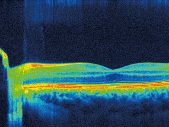
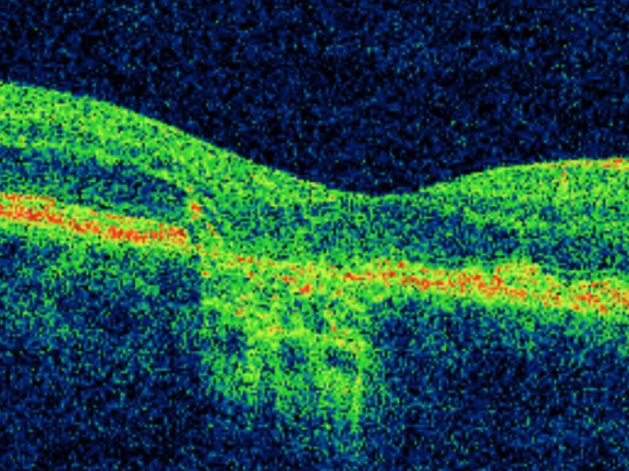
The use of new technologies such as FAF, scanning laser ophthalmoscopes and OCT will be discussed alongside more traditional approaches of Binocular indirect ophthalmoscopy and field testing.
CPD Points: 1
CPDpoints.com credits: 1
Expiry Date: 31/12/2024